Alpha vs Beta Hemolysis: Understanding the Critical Differences
When it comes to identifying bacterial strains in the lab, understanding alpha and beta hemolysis becomes incredibly important. These processes show us how different bacteria interact with red blood cells on blood agar plates - and trust me, this information can be life-changing for proper diagnosis and treatment decisions!
Let me share something fascinating I learned during my microbiology studies: many people think all bacteria behave the same way with red blood cells, but that's far from true. Some bacteria are quite gentle - causing only partial breakdown (alpha hemolysis) while others are more aggressive, causing complete destruction of red blood cells (beta hemolysis).
In this comprehensive guide, I'll walk you through everything you need to know about these two types of hemolysis. We'll explore how they work, what causes them, and why this knowledge is crucial for medical professionals.
What Exactly Is Hemolysis?
Before diving into the differences, let's quickly cover what hemolysis means. Simply put, it's the breakdown of red blood cells. When bacteria attack these cells, they can cause different levels of damage - and that's where our two main types come in.
Think of red blood cells like balloons. Alpha hemolysis is like letting a bit of air out slowly, while beta hemolysis is like popping the balloon completely! This analogy helps students remember the fundamental difference.
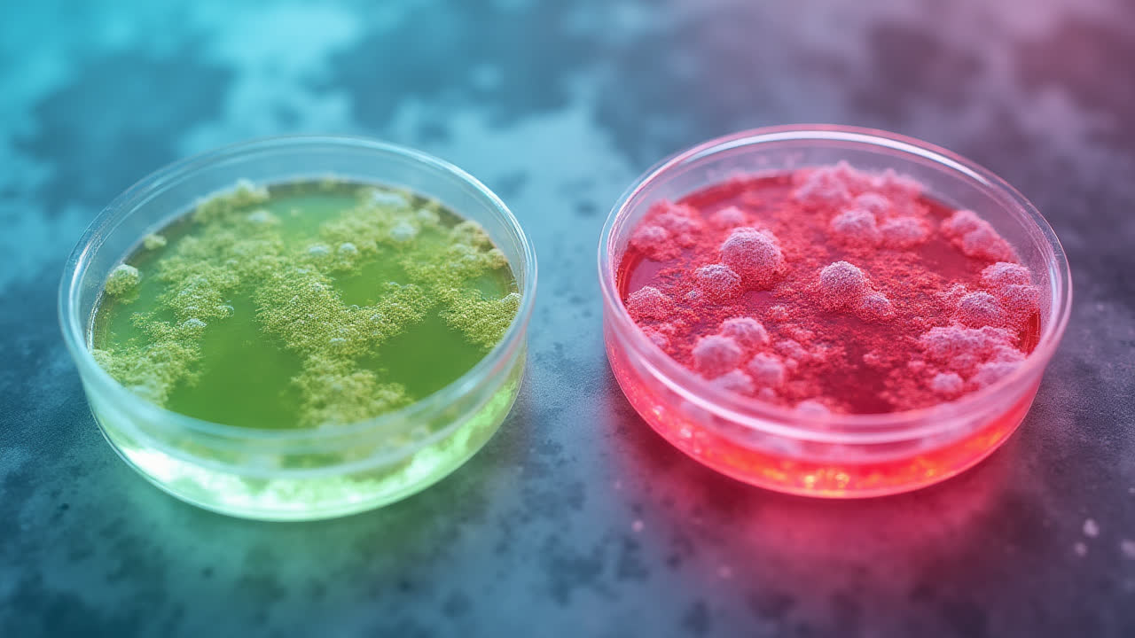
In laboratory settings, we observe these processes on blood agar plates. The visual changes around bacterial colonies tell us exactly what type of hemolysis is occurring. It's quite remarkable how these patterns can help identify specific bacterial species.
Understanding Alpha Hemolysis
Alpha hemolysis is characterized by partial breakdown of red blood cells, creating a distinctive greenish discoloration around bacterial colonies on blood agar. This isn't a complete destruction - rather, it's a transformation of the hemoglobin present in red blood cells.
Here's what happens on a molecular level: bacteria produce hydrogen peroxide, which oxidizes the iron in hemoglobin (red) to methemoglobin (green). This chemical change creates that characteristic greenish-black zone, typically 1-2 mm wide.
The interesting part? Red blood cells remain intact during this process. They're not ruptured or destroyed completely - just modified. This is why we sometimes call it "partial hemolysis."
Common bacteria causing alpha hemolysis include Streptococcus pneumoniae and Streptococcus viridans. These species are often found in the oral cavity and can be important indicators for dental and respiratory infections.
Exploring Beta Hemolysis
Beta hemolysis represents a completely different story - it's the complete breakdown of red blood cells. When you look at a blood agar plate, you'll see a clear zone around the bacterial colony, typically 2-4 mm wide. No greenish tinge here - just transparent areas where all the red blood cells have been destroyed.
The mechanism is fascinating: certain bacteria produce hemolysins like Streptolysin O and Streptolysin S. These powerful toxins literally rupture the cell membranes of red blood cells. It's like a microscopic demolition crew at work!
Streptococcus pyogenes is the classic example of a beta-hemolytic bacterium. Found in the throat, this species is responsible for strep throat and other serious infections. The complete hemolysis helps us identify it quickly in lab settings.
Interestingly, some beta hemolysis reactions can be quite subtle. When dealing with species like Streptococcus agalactiae, you might need to look closely to spot the clear zones. This variability keeps microbiologists on their toes!
Key Differences Between Alpha and Beta Hemolysis
| Feature | Alpha Hemolysis | Beta Hemolysis |
|---|---|---|
| Type of Hemolysis | Partial (incomplete) | Complete |
| Visual Appearance | Greenish discoloration | Clear, transparent zone |
| Red Blood Cell Fate | Remain intact | Completely ruptured |
| Zone Width | 1-2 mm | 2-4 mm |
| Cause | Hydrogen peroxide oxidation | Hemolysin toxins |
| Common Bacteria | S. pneumoniae, S. viridans | S. pyogenes |
| Typical Location | Oral cavity | Throat |
| Chemical Process | Hemoglobin → Methemoglobin | Cell membrane rupture |
Practical Applications in Medical Diagnosis
Understanding these hemolysis patterns isn't just academic - it has real clinical significance. When I worked as a clinical microbiologist, identifying hemolysis patterns was often the first step in determining which antibiotics would be effective against an infection.
For instance, knowing that a patient has a beta-hemolytic streptococcal infection immediately tells us we're likely dealing with S. pyogenes. This information guides treatment decisions and helps predict potential complications.
Laboratories rely on blood agar plates specifically because different hemolysis patterns provide quick, visual identification tools. It's remarkable how a simple color change can tell us so much about the bacteria we're dealing with.
Sometimes, we need to use additional tests to distinguish between similar-looking bacteria. But hemolysis patterns give us that crucial first indication that helps narrow down the possibilities.
Laboratory Techniques and Observations
In the lab, observing hemolysis requires proper technique. Blood agar plates must be prepared correctly, and incubation conditions are crucial. I've seen many students get confused initially because they didn't realize that factors like oxygen levels can affect the results.
For beta hemolysis, creating anaerobic pockets on the agar plate can reveal hemolysis that might not be visible under normal conditions. This technique involves stabbing the agar vertically, which demonstrates how careful methodology impacts results.
Here's a pro tip: always observe hemolysis patterns under proper lighting. What looks like alpha hemolysis in poor lighting might actually be gamma hemolysis (no hemolysis at all). Precision matters in microbiology!
Common Misconceptions and Clarifications
One myth I often encounter is that alpha hemolysis always indicates less pathogenic bacteria. That's not true at all! Streptococcus pneumoniae, which causes pneumonia, shows alpha hemolysis. The type of hemolysis doesn't directly correlate with virulence.
Another misconception is confusing gamma hemolysis (no hemolysis) with alpha hemolysis. Remember, gamma hemolysis appears as the normal red color of blood around the bacterial colony, while alpha hemolysis shows that distinctive green tinge.
Some people think hemolysis patterns are fixed for each bacterial species. In reality, environmental factors, growth conditions, and even strain variations can sometimes affect the patterns observed.
Frequently Asked Questions
What is the main visual difference between alpha and beta hemolysis?
Alpha hemolysis produces a greenish discoloration around the bacterial colony on blood agar, while beta hemolysis creates a clear, transparent zone. Alpha hemolysis appears green because hemoglobin is converted to methemoglobin, whereas beta hemolysis shows complete transparency due to total red blood cell destruction.
Which type of hemolysis is more concerning from a clinical perspective?
Beta hemolysis is often more concerning because it's associated with more aggressive bacteria like S. pyogenes, which can cause serious infections including scarlet fever and rheumatic fever. However, alpha-hemolytic bacteria like S. pneumoniae can also cause severe diseases such as pneumonia and meningitis.
Can environmental factors affect hemolysis patterns?
Yes, environmental factors significantly impact hemolysis patterns. Oxygen levels, temperature, incubation time, and agar composition can all affect the observed hemolysis. For example, Streptolysin O from S. pyogenes is only active in low oxygen conditions, which is why creating anaerobic pockets is important for proper observation.
Conclusion
Understanding the differences between alpha and beta hemolysis is fundamental for anyone working in microbiology or medicine. These patterns provide valuable clues for bacterial identification and guide treatment decisions. While alpha hemolysis shows partial breakdown with that characteristic green color, beta hemolysis demonstrates complete destruction with clear zones.
Remember, these patterns aren't just academic curiosities - they're practical tools that help medical professionals save lives. Whether you're a student learning about bacterial identification or a healthcare worker interpreting lab results, this knowledge forms a cornerstone of microbiological diagnosis.
As you continue your journey in understanding bacterial hemolysis, keep observing, questioning, and learning. The microscopic world has many more secrets to reveal, and every pattern tells a story worth understanding.